Showing posts with label anatomy. Show all posts

Friday, September 03, 2010

Diaphragm


The large muscle primarily responsible for inhalation is the?
Diaphragm...also help to stabilize the trunk


Is the diaphragm muscle skeletal muscle?
The diaphragm is a skeletal muscle that separates the thoracic and abdominopelvic cavities. It is innervated by the phrenic nerve. The diaphragm is the main muscle of respiration, with diaphragmatic...

Monday, May 24, 2010

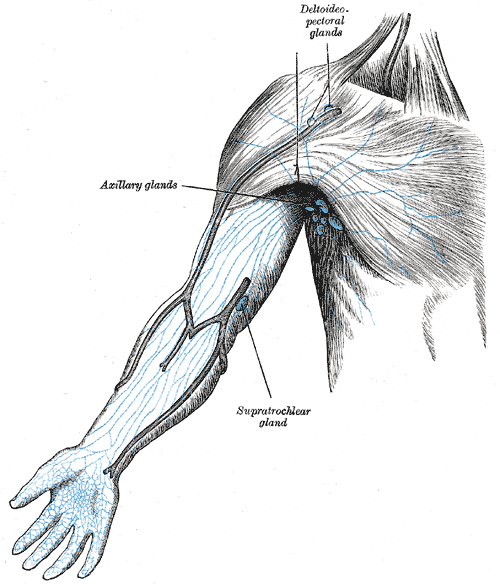
Nodus Limfa


Kebanyakkan manusia tidak menyedari kepentingan nodus limfa dlm tubuh badan mereka. Sy juga begitu tapi bagi pesakit2 yg kehilangan salah satu nodus limfa mereka, mereka memahami kepentingan tersebut. Sy jg perlu tahu kepentingannya kerana sy perlu memberi penjelasan kpd pesakit tntg penjagaan diri slps pembedahan...huhhu,skema btul ayat2ku ini..=P Antara kes2 yg melibatkan pembuangan nodus limfa ialah kanser payudara, kanser servik("hysterectomy, (ophorectomy Mastektomi....post ni xsiap lg sebenarnya..hohoho..aigOo


Nodus limfa adalah salah satu komponen dari sistem limfatik yang dapat ditemukan pada tubuh manusia. Nodus limfa adalah filter untuk partikel asing dan berisi sel darah putih. Nodus limfa berfungsi sebagai filter, dengan jaringan konektif retikular dan limfosit yang mengumpulkan dan menghancurkan bakteri dan virus. Ketika tubuh melawan infeksi, limfosit bertambah dengan deras.

Wednesday, May 12, 2010

Dermatome


Dermatome is a Greek word which literally means "skin cutting". A dermatome is an area of the skin supplied by nerve fibers originating from a single dorsal nerve root. The dermatomes are named according to the spinal nerve which supplies them. The dermatomes form into bands around the trunk but in the limbs their organisation is more complex as a result of the dermatomes being "pulled out" as the limb buds form and develop into the limbs during embryological development.

In diagrams or maps, the boundaries of dermatomes are usually sharply defined. However, in life there is considerable overlap of innervation between adjacent dermatomes. Thus, if there is a loss of afferent nerve function by one spinal nerve sensation from the region of skin which it supplies is not usually completely lost as overlap from adjacent spinal nerves occurs: however, there will be a reduction in sensitivity.

Myotome


Each muscle in the body is supplied by a particular level or segment of the spinal cord and by its corresponding spinal nerve. The muscle, and its nerve make up a myotome. This is approximately the same for every person and are as follows:

C3,4 and 5 supply the diaphragm (the large muscle between the chest and the belly that we use to breath).

C5 also supplies the shoulder muscles and the muscle that we use to bend our elbow .

C6 is for bending the wrist back.

C7 is for straightening the elbow.

C8 bends the fingers.

T1 spreads the fingers.

T1 –T12 supplies the chest wall & abdominal muscles.

L2 bends the hip.

L3 straightens the knee.

L4 pulls the foot up.

L5 wiggles the toes.


S1 pulls the foot down.

S3,4 and 5 supply the bladder. bowel and sex organs and the anal and other pelvic muscles.

Quadriceps muscle


The Quadriceps are a group of four muscles that sit on the anterior or front aspect of the thigh.


Rectus Femoris:
Origin:
- straight head: anterior inferior iliac spine
- reflected head: groove on upper brim of acetabulum
Insertion: upper border of patella by the ligamentum patella into tibia tuberosity
Reversed origin-insertion action: flexes the pelvis on the femur and gives anterior stabilization to the pelvis
Nerve supply: femoral, L2, L3, L4
Synergists: psoas, sartorius, tensor fascia lata, vastus lateralis, vastus medialis, vastus intermedius;
Arterial supply: ascending branch of LFCA
Ref: Contribution of rectus femoris and vasti to knee extension. An electromyographic study.

Vastus Intermedius:
Origin: proximal 2/3 of the anterolateral surface of the femur, lower 1/2 of the linea aspera, upper part of the lateral supracondylar line; lateral intermuscular septum
Insertion: by tendons of the rectus and vastus muscles into the superior border of the patella and by the ligamentum patella into the tibial tuberosity
Action: extends the leg at the knee
Nerve supply: femoral, L2, L3, L4
Synergists: rectus femoris, vastus medialis, vastus lateralis
Arterial supply: descending branch of LFCA

Vastus Lateralis:
Origin: upper part of intertrochanteric line, anterior and lower borders of greater trochanter, lateral lip of gluteal tuberosity, upper half of linea aspera, lateral intermuscular septum, and tendon of maximus
Insertion: lateral border of the patella by the ligamentum patella into the tibia tubercle;
Action: extends the leg at the knee and draws the patella laterally;
Nerve supply: femoral, L3 > L2, L4; (see innervation)
Synergists: rectus femoris, vastus intermedius, vastus medialis;
Arterial supply: descending and transverse brs. of LFCA;

Vastus Medialis:
Origin: lower 1/2 of the intertrochanteric lines, medial lip of linea aspera, upper part of medial supracondylar line, medial intermuscular septum tendons of adductor magnus and adductor longus
Insertion: Medial border of the patella by the ligamentum patella into the tibia tuberosity
Action: extends the leg at the knee and draws the patella medially;
Nerve Supply: femoral, L3 > L2, L4; (See innervation)
Synergists: rectus femoris, vastus lateralis, vastus intermedius


Gait Characteristics:
- at normal walking speeds the limb behaves as pendulum, w/ near full extension of the knee joint as it preparare for heel-strike
- during the first 5% of the gait cycle, the quadcrips generate peak intensity of 25%
- after the first 20% of the gait cycle, the quadriceps become silent
- exam:
- note that normally an examiner will not be able to "break" a quadriceps unless it is severely weakened
- 4/5 strength will have 40% of normal strength
- note that even w/ +3 stength, gait will be relatively normal

Quad Paralysis:
- when quadriceps muscle is paralyzed, walking on level surface may appear to be entirely normal
- at full extension, action of the quadriceps is not necessary for stability of knee joint; if the line of gravity is maintained anterior to the axis of motion of the knee joint, full extension persists through the stance phase;
- w/ quad paralysis, however, the patient will be unable to run and may have difficulty with stairs, because in these cases, full extension is not attained and the knee tends to buckle into flexion
- patients will often walk with the leg externally rotated inorder to help "lock" the knee at heel strike;
Associated conditions:
- in the case of a weak quadriceps and triceps, the occurance of an equinus contracture or a hinged AFO w/ dorsiflexion block will both prevent excessive knee flexion (buckling) and excessive ankle dorsiflexion during stance phase;
- hence, in this case the equinus contracture is beneficial
Management:
- when this muscle is quite weak, a long leg brace may be needed to support knee joint in full extension, inorder to prevent knee from buckling during gait
- consider adding a cushioned heel to the patient's shoe inorder to provide additional knee stability at heel-strikeheel strike

Showing posts with label anatomy. Show all posts

Friday, September 03, 2010

Diaphragm


The large muscle primarily responsible for inhalation is the?
Diaphragm...also help to stabilize the trunk


Is the diaphragm muscle skeletal muscle?
The diaphragm is a skeletal muscle that separates the thoracic and abdominopelvic cavities. It is innervated by the phrenic nerve. The diaphragm is the main muscle of respiration, with diaphragmatic...

Monday, May 24, 2010

Nodus Limfa


Kebanyakkan manusia tidak menyedari kepentingan nodus limfa dlm tubuh badan mereka. Sy juga begitu tapi bagi pesakit2 yg kehilangan salah satu nodus limfa mereka, mereka memahami kepentingan tersebut. Sy jg perlu tahu kepentingannya kerana sy perlu memberi penjelasan kpd pesakit tntg penjagaan diri slps pembedahan...huhhu,skema btul ayat2ku ini..=P Antara kes2 yg melibatkan pembuangan nodus limfa ialah kanser payudara, kanser servik("hysterectomy, (ophorectomy Mastektomi....post ni xsiap lg sebenarnya..hohoho..aigOo


Nodus limfa adalah salah satu komponen dari sistem limfatik yang dapat ditemukan pada tubuh manusia. Nodus limfa adalah filter untuk partikel asing dan berisi sel darah putih. Nodus limfa berfungsi sebagai filter, dengan jaringan konektif retikular dan limfosit yang mengumpulkan dan menghancurkan bakteri dan virus. Ketika tubuh melawan infeksi, limfosit bertambah dengan deras.

Wednesday, May 12, 2010

Dermatome


Dermatome is a Greek word which literally means "skin cutting". A dermatome is an area of the skin supplied by nerve fibers originating from a single dorsal nerve root. The dermatomes are named according to the spinal nerve which supplies them. The dermatomes form into bands around the trunk but in the limbs their organisation is more complex as a result of the dermatomes being "pulled out" as the limb buds form and develop into the limbs during embryological development.

In diagrams or maps, the boundaries of dermatomes are usually sharply defined. However, in life there is considerable overlap of innervation between adjacent dermatomes. Thus, if there is a loss of afferent nerve function by one spinal nerve sensation from the region of skin which it supplies is not usually completely lost as overlap from adjacent spinal nerves occurs: however, there will be a reduction in sensitivity.

Myotome


Each muscle in the body is supplied by a particular level or segment of the spinal cord and by its corresponding spinal nerve. The muscle, and its nerve make up a myotome. This is approximately the same for every person and are as follows:

C3,4 and 5 supply the diaphragm (the large muscle between the chest and the belly that we use to breath).

C5 also supplies the shoulder muscles and the muscle that we use to bend our elbow .

C6 is for bending the wrist back.

C7 is for straightening the elbow.

C8 bends the fingers.

T1 spreads the fingers.

T1 –T12 supplies the chest wall & abdominal muscles.

L2 bends the hip.

L3 straightens the knee.

L4 pulls the foot up.

L5 wiggles the toes.


S1 pulls the foot down.

S3,4 and 5 supply the bladder. bowel and sex organs and the anal and other pelvic muscles.

Quadriceps muscle


The Quadriceps are a group of four muscles that sit on the anterior or front aspect of the thigh.


Rectus Femoris:
Origin:
- straight head: anterior inferior iliac spine
- reflected head: groove on upper brim of acetabulum
Insertion: upper border of patella by the ligamentum patella into tibia tuberosity
Reversed origin-insertion action: flexes the pelvis on the femur and gives anterior stabilization to the pelvis
Nerve supply: femoral, L2, L3, L4
Synergists: psoas, sartorius, tensor fascia lata, vastus lateralis, vastus medialis, vastus intermedius;
Arterial supply: ascending branch of LFCA
Ref: Contribution of rectus femoris and vasti to knee extension. An electromyographic study.

Vastus Intermedius:
Origin: proximal 2/3 of the anterolateral surface of the femur, lower 1/2 of the linea aspera, upper part of the lateral supracondylar line; lateral intermuscular septum
Insertion: by tendons of the rectus and vastus muscles into the superior border of the patella and by the ligamentum patella into the tibial tuberosity
Action: extends the leg at the knee
Nerve supply: femoral, L2, L3, L4
Synergists: rectus femoris, vastus medialis, vastus lateralis
Arterial supply: descending branch of LFCA

Vastus Lateralis:
Origin: upper part of intertrochanteric line, anterior and lower borders of greater trochanter, lateral lip of gluteal tuberosity, upper half of linea aspera, lateral intermuscular septum, and tendon of maximus
Insertion: lateral border of the patella by the ligamentum patella into the tibia tubercle;
Action: extends the leg at the knee and draws the patella laterally;
Nerve supply: femoral, L3 > L2, L4; (see innervation)
Synergists: rectus femoris, vastus intermedius, vastus medialis;
Arterial supply: descending and transverse brs. of LFCA;

Vastus Medialis:
Origin: lower 1/2 of the intertrochanteric lines, medial lip of linea aspera, upper part of medial supracondylar line, medial intermuscular septum tendons of adductor magnus and adductor longus
Insertion: Medial border of the patella by the ligamentum patella into the tibia tuberosity
Action: extends the leg at the knee and draws the patella medially;
Nerve Supply: femoral, L3 > L2, L4; (See innervation)
Synergists: rectus femoris, vastus lateralis, vastus intermedius


Gait Characteristics:
- at normal walking speeds the limb behaves as pendulum, w/ near full extension of the knee joint as it preparare for heel-strike
- during the first 5% of the gait cycle, the quadcrips generate peak intensity of 25%
- after the first 20% of the gait cycle, the quadriceps become silent
- exam:
- note that normally an examiner will not be able to "break" a quadriceps unless it is severely weakened
- 4/5 strength will have 40% of normal strength
- note that even w/ +3 stength, gait will be relatively normal

Quad Paralysis:
- when quadriceps muscle is paralyzed, walking on level surface may appear to be entirely normal
- at full extension, action of the quadriceps is not necessary for stability of knee joint; if the line of gravity is maintained anterior to the axis of motion of the knee joint, full extension persists through the stance phase;
- w/ quad paralysis, however, the patient will be unable to run and may have difficulty with stairs, because in these cases, full extension is not attained and the knee tends to buckle into flexion
- patients will often walk with the leg externally rotated inorder to help "lock" the knee at heel strike;
Associated conditions:
- in the case of a weak quadriceps and triceps, the occurance of an equinus contracture or a hinged AFO w/ dorsiflexion block will both prevent excessive knee flexion (buckling) and excessive ankle dorsiflexion during stance phase;
- hence, in this case the equinus contracture is beneficial
Management:
- when this muscle is quite weak, a long leg brace may be needed to support knee joint in full extension, inorder to prevent knee from buckling during gait
- consider adding a cushioned heel to the patient's shoe inorder to provide additional knee stability at heel-strikeheel strike